As we age, it is natural for our bodies to undergo changes, and this includes our eyesight. One of the most common issues that seniors face is macular degeneration, a condition that affects the central area of the retina, which is responsible for seeing details and colors.
There are two types of macular degeneration, dry and wet, and while the former is more common, the latter is more severe.
Let’s explore wet macular degeneration in detail, including the causes, symptoms, and available treatment options.
What Is Wet Macular Degeneration?
When the blood vessels in the eye that feed oxygen and nutrients to the retina grow abnormally, it leads to wet macular degeneration.
These abnormal blood vessels leak fluid, causing swelling and damage to the macula, the central part of the retina responsible for sharp, clear vision. This condition can lead to central vision loss, making it difficult to read, recognize faces, drive, or perform other activities that require sharp vision.
Although less common than its counterpart, dry macular degeneration, wet macular degeneration accounts for approximately 90% of all severe vision loss from the disease.
There are two primary ways that wet macular degeneration can lead to vision loss:
Irregular Blood Vessel Growth
This condition, known as choroidal neovascularization, involves the growth of abnormal blood vessels from the choroid (a layer of blood vessels between the retina and the outer coat of the eye called the sclera) under and into the macula (the part of the eye responsible for sharp, central vision).
These abnormal blood vessels can leak fluid or blood, disrupting the functioning of the retina and leading to vision loss.
Fluid Buildup in the Back of the Eye
Fluid leaking from the choroid can accumulate between the retinal pigment epithelium (a thin cell layer) and the retina or within the layers of the retina itself. This can cause irregularities in the macula layers, resulting in loss of vision or distorted vision.
Symptoms of Wet Macular Degeneration
The symptoms of wet macular degeneration can vary and may develop suddenly and quickly.
Some of the most common symptoms include:
- Seeing straight lines as wavy
- Sudden changes in vision
- Blind spots in the central vision
- Difficulty seeing in low-light conditions
- Difficulty recognizing faces
If you experience any of these symptoms, especially changes in your central vision or inability to see fine detail, it is important to seek medical attention promptly to prevent the condition from worsening.
Causes of Wet Macular Degeneration
The exact cause of wet macular degeneration is unknown, but there are several factors that are believed to contribute to the development of the condition. These include genetic predisposition, smoking, high blood pressure, obesity, and a diet low in vitamins (especially C and E) and minerals (copper and zinc). It is also more common in Caucasians.
If you have a family history of macular degeneration or any of these risk factors, it is important to take steps to prevent or manage the condition.
Diagnosis of Wet Macular Degeneration
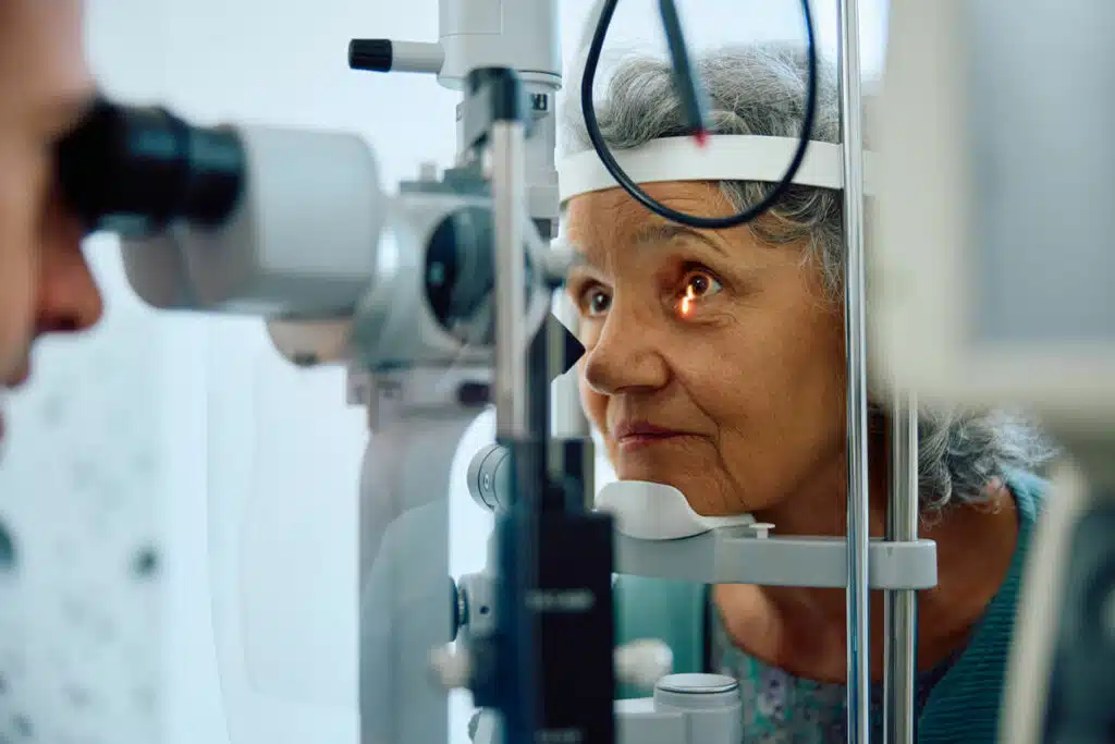
To diagnose wet macular degeneration, an ophthalmologist will conduct a comprehensive eye exam. This may involve a dilated eyes exam and other tests, including:
- Fluorescein Angiography: This test involves injecting a dye into a vein in your arm, which travels to and highlights the blood vessels in your eye. A special camera captures images as the dye circulates through the blood vessels. These images can reveal any leaking blood vessels or retinal changes.
- Indocyanine Green Angiography: Similar to fluorescein angiography, this test also uses an injected dye. It may be used to confirm the findings of a fluorescein angiography or to identify problematic blood vessels located deeper in the retina.
- Optical Coherence Tomography (OCT): OCT is a non-invasive imaging test that provides detailed cross-sections of the retina. It can identify areas of thinning, thickening, or swelling in the retina.
Additionally, OCT is used to monitor how the retina responds to macular degeneration treatments.
- Optical Coherence Tomography Angiography (OCTA): OCTA is a newer, non-invasive test that, in certain cases, allows ophthalmologists to see unwanted blood vessels in the macula. While it is primarily used as a research tool, its clinical use is increasing.
Treatment Options
While there is no cure for wet macular degeneration, several treatment options can help slow its progression. Common wet macular degeneration treatments include:
Anti-VEGF Therapy
Anti-VEGF drugs are designed to block a protein called vascular endothelial growth factor (VEGF), which stimulates the growth of new blood vessels. These drugs are injected directly into the eye to halt the growth of abnormal blood vessels and prevent further leakage.
Drugs are injected every 4 to 6 weeks to maintain their effect.
Photodynamic Therapy (PDT)
PDT involves injecting a light-sensitive drug (verteporfin) into the bloodstream, which gets absorbed by the abnormal blood vessels in the eye. A laser is then used to activate the drug, damaging the abnormal blood vessels without harming the surrounding tissue.
Laser Surgery
In some cases, a high-energy laser beam may be used to destroy abnormal blood vessels.
Wet Macular Degeneration Treatment Near Me
If you or a loved one are experiencing symptoms of wet macular degeneration, we urge you to schedule an appointment with us today at Advanced Sight Center. Our dedicated team of professionals specializes in macular degeneration treatments and is committed to providing you with the highest level of care.
To know more about us or to schedule a consultation, call us today at (636) 239-1650 or fill out our online appointment request form. We look forward to serving you!


